Understanding Knee Anatomy: A Comprehensive Guide
Introduction
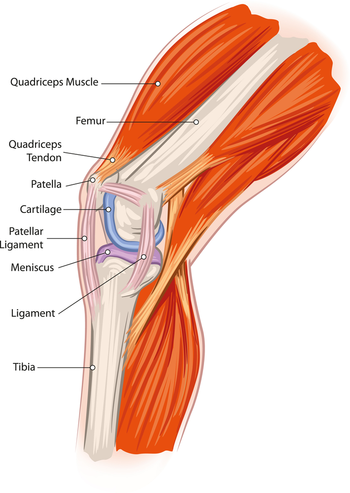
The knee is one of the most complex and crucial joints in the human body. Understanding its anatomy is essential for diagnosing and treating various knee-related issues. This article delves into the detailed anatomy of the knee, including its bones, ligaments, tendons, muscles, and cartilage, providing a comprehensive overview for anyone interested in this vital joint.
Bones of the Knee
The knee joint comprises three primary bones: the femur (thighbone), the tibia (shinbone), and the patella (kneecap). These bones work together to facilitate movement and support body weight.
Femur
The femur is the longest and strongest bone in the human body. At the knee joint, the femur’s distal end forms two rounded protrusions called condyles, which articulate with the tibia.
Tibia
The tibia, or shinbone, is the larger and stronger of the two bones in the lower leg. Its proximal end features two plateaus (medial and lateral) that interact with the femur’s condyles.
Patella
The patella, or kneecap, is a small, triangular bone that protects the knee joint. It sits within the quadriceps tendon and enhances the leverage of the quadriceps muscle, aiding in knee extension.
Ligaments of the Knee
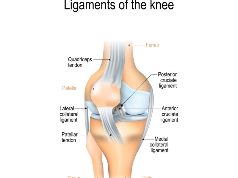
Ligaments are strong, fibrous tissues that connect bones and stabilize joints. The knee contains several critical ligaments.
Anterior Cruciate Ligament (ACL)
The ACL runs diagonally in the middle of the knee, preventing the tibia from sliding out in front of the femur and providing rotational stability.
Posterior Cruciate Ligament (PCL)
The PCL is stronger than the ACL and prevents the tibia from sliding backward under the femur. It also provides stability, particularly during movements involving bending.
Medial Collateral Ligament (MCL)
The MCL runs along the inside of the knee, connecting the femur to the tibia. It resists valgus forces, which push the knee inward.
Lateral Collateral Ligament (LCL)
The LCL is located on the outer side of the knee, connecting the femur to the fibula. It resists varus forces, which push the knee outward.
Tendons of the Knee
Tendons are fibrous tissues that connect muscles to bones. The knee has several important tendons that contribute to its movement and stability.
Quadriceps Tendon
The quadriceps tendon connects the quadriceps muscle group to the patella. It plays a crucial role in extending the knee.
Patellar Tendon
The patellar tendon, technically a ligament, connects the patella to the tibia. It is essential for transmitting the force generated by the quadriceps muscle to the lower leg.
Hamstring Tendons
The hamstring tendons, including the semitendinosus, semimembranosus, and biceps femoris tendons, attach the hamstring muscles to the pelvis, femur, and tibia, allowing knee flexion and hip extension.
Muscles of the Knee
The knee’s movement and stability depend on various muscle groups.
Quadriceps
The quadriceps are a group of four muscles on the front of the thigh: the rectus femoris, vastus lateralis, vastus medialis, and vastus intermedius. These muscles are responsible for knee extension.
Hamstrings
The hamstrings are located at the back of the thigh and include the biceps femoris, semitendinosus, and semimembranosus. They facilitate knee flexion and hip extension.
Calf Muscles
The gastrocnemius and soleus muscles form the calf. These muscles contribute to knee flexion and are involved in walking, running, and jumping.
Cartilage of the Knee
Cartilage is a smooth, rubbery tissue that covers and protects the ends of bones, allowing smooth joint movement.
Articular Cartilage
Articular cartilage covers the ends of the femur, tibia, and patella. It reduces friction during movement and absorbs shock.
Menisci
The menisci are two C-shaped pieces of fibrocartilage located between the femur and tibia. They act as shock absorbers, enhance joint stability, and distribute weight evenly across the knee.
Conclusion
The knee is a remarkably complex joint, essential for everyday movements and athletic activities. Its intricate anatomy, comprising bones, ligaments, tendons, muscles, and cartilage, works together to provide stability, flexibility, and strength. Understanding the knee’s anatomy is crucial for diagnosing injuries, planning treatments, and implementing effective rehabilitation strategies. Whether you’re an athlete, healthcare professional, or someone interested in human anatomy, knowing the details of knee anatomy can significantly enhance your comprehension of this vital joint.

